3 D LIMON Super Resolution Microscopy
3 D LIMON (LightMicrOscopical nanosizing microscopy) images using the Vertico SMI microscope are made possible by the combination of SMI and SPDM, whereby first the SMI and then the SPDM process is applied.
The SMI process determines the center of particles and their spread in the direction of the microscope axis. While the center of particles/molecules can be determined with a 1–2 nm precision, the spread around this point can be determined down to an axial diameter of approx. 30–40 nm. Subsequently, the lateral position of the individual particle/molecule is determined using SPDM, achieving a precision of a few nanometers.
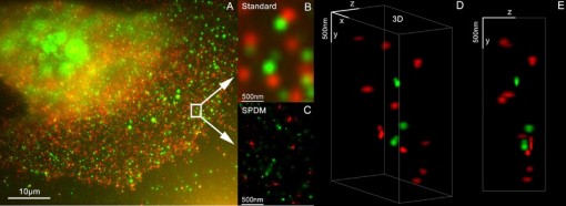
As a biological application in the 3D dual color mode the spatial arrangements of Her2/neu and Her3 clusters was achieved. The positions in all three directions of the protein clusters could be determined with an accuracy of about 25 nm.
References
- Baddeley D, Batram C, Weiland Y, Cremer C, Birk UJ.: Nanostructure analysis using Spatially Modulated Illumination microscopy. In: Nature protocols 2007; 2: 2640–2646
- Rainer Kaufmann, Patrick Müller,Georg Hildenbrand, Michael Hausmann & Christoph Cremer (2010): Analysis of Her2/neu membrane protein clusters in different types of breast cancer cells using localization microscopy, Journal of Microscopy 2010